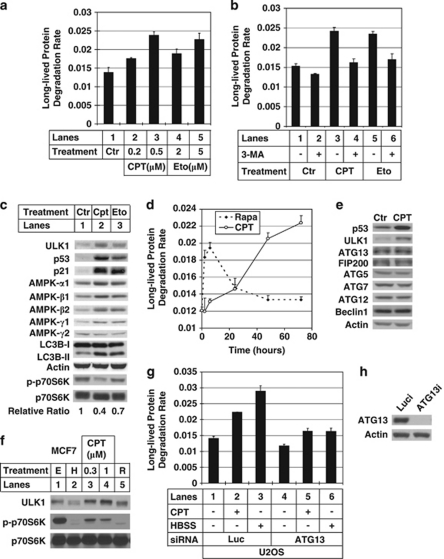
Figure 1.
Autophagy induction by sub-lethal DNA damage. (a) U2OS cells were treated with different concentrations of camptothecin (CPT, lanes 2 and 3) or etoposide (Eto, lanes 4 and 5) for 2 days before doing the long-lived protein degradation (LLPD) assay as described in Materials and Methods. Y axis is the LLPD rate after 2 h. (b) U2OS cells were treated with either 0.5 μM CPT (lanes 3 and 4) or 5 μM Eto (lanes 5 and 6) for 2 days. On the day of measuring LLPD rate, 5 mM of 3-methyladenine (3-MA) was used to pretreat cells for 1 h (lanes 2, 4 and 6) before doing the LLPD assay. (c) U2OS cells were treated with either 0.5 μM CPT (lane 2) or 5 μM Eto (lane 3) for 2 days. The cells were collected and lysed to run western blot. The blot were probed with the antibody against unc-51-like kinase 1 (ULK1), p53, p21, T-389 phosphorylated form of p70S6K (p-p70S6K), p70S6K, AMPK-α1, -β1, -β2, -γ1, -γ2, LC3B and actin. To measure the ratio of p-p70S6K over p70S6K, the blot was developed using enhanced chemiluminescence (ECL) reagent and the band intensities were measured using Kodak Image Station 4000R pro. The relative ratio for the control is defined as 1. (d) U2OS cells were treated with 1 μM rapamycin or 0.5 μM CPT for 2, 6, 24, 48 and 72 h, after which LLPD rates were measured for each sample and plotted against treatment time. (e) U2OS cells were treated with 1 μM CPT for 1 day. The cells were then collected and lysed to do western blot. The blots were probed with the antibodies against p53, ULK1, ATG13, FIP200, ATG5, ATG7, ATG12, beclin 1 and actin. (f) MCF7 cells were untreated (E, lane 1) or treated with either Hank's buffered salt solution (HBSS) for 2 h (H, lane 2), 0.3 μM CPT for 1 day (lane 3), 1 μM CPT for 1 day (lane 4) and 1 μM rapamycin for 6 h (R, lane 5). The cells were then collected and lysed to do western blot. The blots were probed with the antibodies against ULK1, p-p70S6K and p70S6K. (g) U2OS cells were transfected with either luciferase control small interfering (si)RNA (lanes 1–3) or ATG13 siRNA (lanes 4–6) as described in Materials and Methods. After 3 days of siRNA transfection, they were treated with (lanes 2 and 5) or without (lanes 1 and 4) 0.5 μM CPT for 2 days and measured the LLPD rates. HBSS treatments (lanes 3 and 6) were used as controls. (h) The same samples as in (g) were used to do western blot to confirm the knockdown efficiency of ATG13. These data are presented as mean+S.D. and are representative of at least two independent experiments

