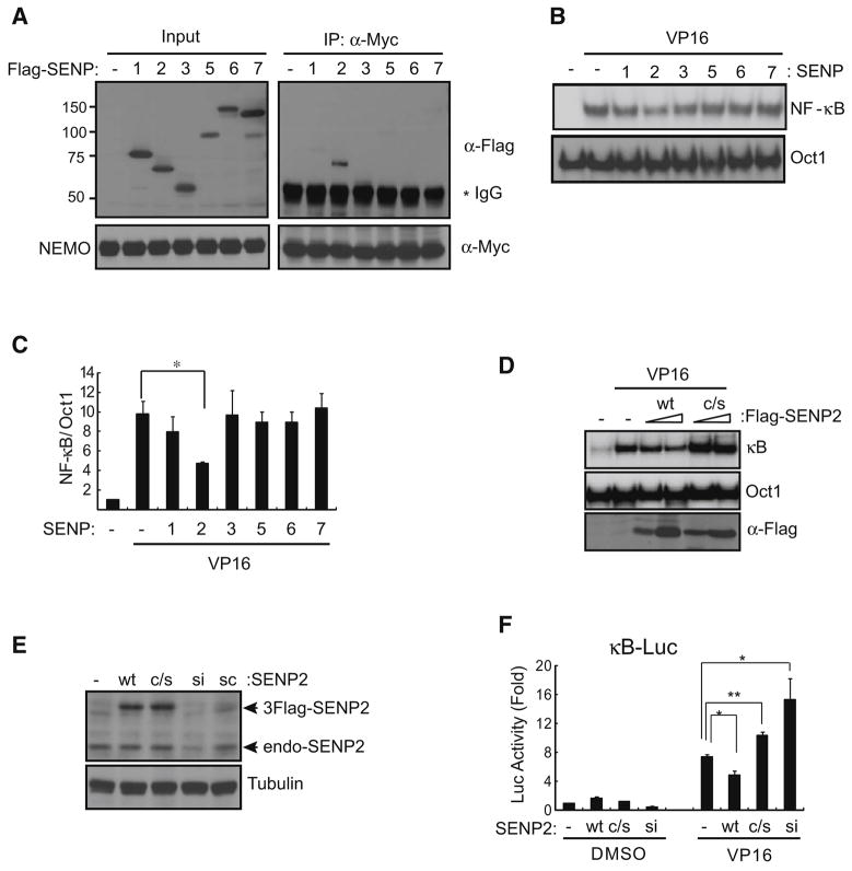
Fig. 1.
SENP2 associates with NEMO and negatively regulates NF-κB activation by genotoxic stress. (A) Flag-SENP1, 2, 3, 5, 6, and 7 were co-transfected with Myc-NEMO in HEK293 cells. Transfected cells were treated with 10 μM VP16 for 1 h, lysed and NEMO was then precipitated with Myc antibody and analyzed by immunoblot using Flag or NEMO antibody. (B) HEK293 cells were transfected with vector (−) or SENP constructs and treated as above. Lysates were used for EMSA using a radio-labeled Igκ-κB or Oct-1 probe. (C) Phosphorimage quantified results from four experiments as in (B) were plotted (mean+SEM). *, p<0.03. (D) HEK293 cells were transfected with varying amounts of Flag-SENP2 wild type (wt) or a catalytically inactive mutant (c/s) and were analyzed by EMSA as in (A). (E) HEK293 cells were co-transfected with SENP2-wt, -c/s, or -siRNA (SMARTpool, Dharmacon) and 3x-κB-luciferase and β-galactosidase constructs. Twenty-four hours following transfection, cells were treated with VP16 or DMSO for 8 h and extracts were analyzed by immunoblotting using SENP2 or tubulin antibody. (F) Cell samples as in (E) were analyzed for luciferase and β-galactosidase activities and relative luciferase activity was plotted (mean+SEM). *, p<0.03; **, p<0.001. See also Figure S1.

