
Figure 6.
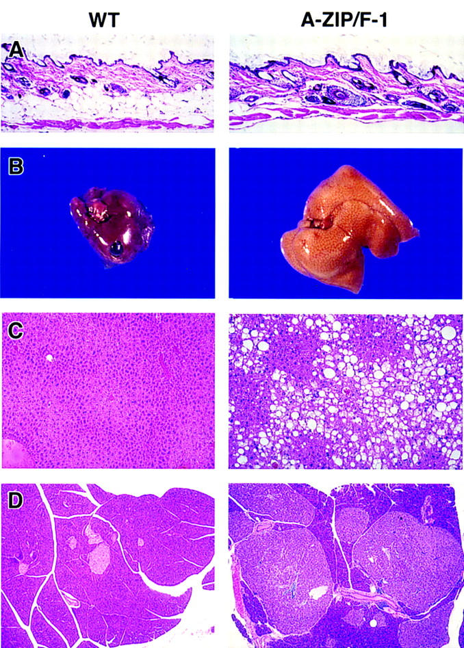
Appearance of skin, liver, and pancreas in mice at 22 weeks of age. Tissues from female wild-type (WT, left) and A-ZIP/F-1 (right) mice are shown at the same magnifications. (A) Skin, with WAT visible below the dermis in wild-type but not A-ZIP/F-1 mice. Grossly, livers of A-ZIP/F-1 mice are enlarged and lighter in color (B). Microscopically the A-ZIP/F-1 liver is filled with lipid droplets (C). Pancreatic islet β cell hypertrophy and hyperplasia were present in all A-ZIP/F-1 mice examined. A particularly striking example is in D (the lighter staining cells are the islets and the darker region is the exocrine pancreas). Hematoxylin and eosin staining magnification, 30× (A,C) and 16× (D).