Figure 2.
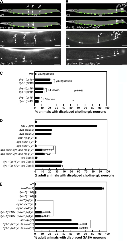
Mutations in dys-1 cause defects in maintaining neuronal positions. (A and B) Schematics and corresponding micrographs of VNC cholinergic neurons, as visualized in a young adult wild-type (WT) and dys-1(cx18) animal expressing the Punc-129::gfp marker (A) and VNC GABA neurons, as visualized in a young adult wild-type and a dys-1(cx18)/+;sax-7(eq1)/+ animal expressing UNC-47::GFP (B). The brackets in A mark the vulval muscles, which also express unc-129::gfp. The green dots and lines represent the neuronal cell bodies and axonal processes, respectively. (C–E) The cholinergic neurons DB5, DA5, and DB6 and GABA neurons VD11, VD12, DD6, and VD13 are stereotypically positioned in wild-type animals (see Materials and methods; Fig. S4). The relative positions of the cholinergic neurons are altered in >20% dys-1 adult animals, a phenotype that is less prevalent in dys-1 larvae (C), suggesting a positional maintenance role for dys-1. The quantification of young adult animals exhibiting displaced cholinergic (D) and GABA (E) neurons in dys-1 and sax-7 mutant backgrounds reveals a genetic interaction between dys-1 and sax-7. Error bars show the standard error of the proportions of three sample sets in which n = 100 in each set. The p-values in C–E show the statistical significance as assessed by the Z test between the indicated strains. Bars, 10 µm.

