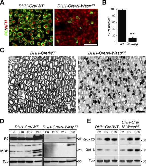
Figure 2.
DHH-Cre/N-Waspfl/fl mice exhibit a severe PNS myelination defect. (A) Transverse sections of P60 sciatic nerves from control and mutant mice immunolabeled with antibodies to P0 and neurofilament (NFH). Bar, 50 µm. (B) There was a strong reduction in the number of axons encircled by a P0-positive myelin sheath in the mutant. Error bars represent the SD of n = 3 mice for each genotype (**, P < 0.0001). (C) Transverse semithin sections of sciatic nerves isolated from control and mutant mice at P30 stained with toluidine blue. Although in the control nerve most axons were enveloped by a mature myelin sheath, only a few thin myelin profiles were detected in the mutant (arrowheads). Bar, 20 µm. (D and E) Western blot analysis of sciatic nerves from control and mutant mice at different ages using the indicated antibodies. Tubulin (Tub) served as a loading control. Molecular masses are given in kilodaltons. WT, wild type.

