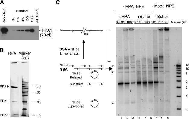
Figure 1.
Effect of RPA depletion on SSA. (A) Western blot of RPA- or mock-depleted NPE. RPA was detected with a rat antibody against the large subunit (p70). The standards for quantitation were extracts loaded at the indicated amounts relative to the depleted extracts. (B) Silver staining of the RPA protein purified from Xenopus egg extracts. 0.5 µg of protein was separated by a 12% SDS-PAGE and then detected by silver staining (Bio-Rad Laboratories). The right lane is the protein size marker (Invitrogen). (C) SSA repair in RPA- and mock-depleted NPE. 12 ng/µl of the pRW4* substrate was incubated in RPA- and mock-depleted NPE at room temperature, and the samples taken at the indicated times were treated with SDS-EDTA–proteinase K, separated on a 1% TAE-agarose gel, and detected by SYBR gold staining. The NHEJ products are marked by the asterisks, and the SSA products are marked by the arrow and the bracket. (n) represents multiple repeats.

