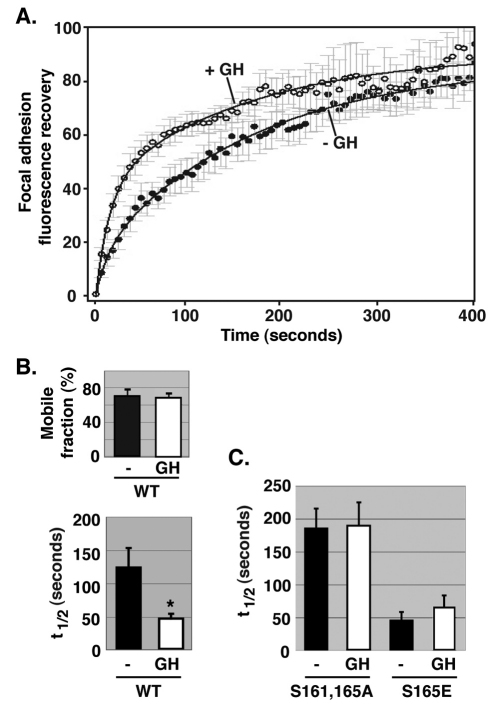
Fig. 6.
Growth hormone regulates the focal adhesion dynamics of SH2B1β. (A) 3T3-F442A cells expressing GFP–SH2B1β were incubated in serum-free medium overnight and then individual focal adhesions were photobleached. During the photobleaching scans, cells were treated with (n=5) or without (n=8) 500 ng/ml GH. FRAP analysis was carried out for 400 seconds. FRAP values were obtained using Olympus Fluoview software. Data were normalized and best-fit curves were determined as described in the Materials and Methods. (B) Mobile fraction and t1/2 values were calculated as described in the Materials and Methods. Error bars indicate s.e.m. *P<0.05 by two-tailed Student's t-test compared with the data from cells treated without GH. (C) 3T3-F442A cells expressing GFP–SH2B1β (S161A, S165A) or GFP–SH2B1β (S165E) were treated and the results were analyzed as in A. t1/2 values were calculated as in B. Error bars indicate s.e.m. (n=3). The t1/2 for GFP–SH2B1β (S165E) (+ GH or − GH) is less than that of GFP–SH2B1β (S161A, S165A) (+ GH or − GH) (P<0.05 by two-tailed Student's t-test).

