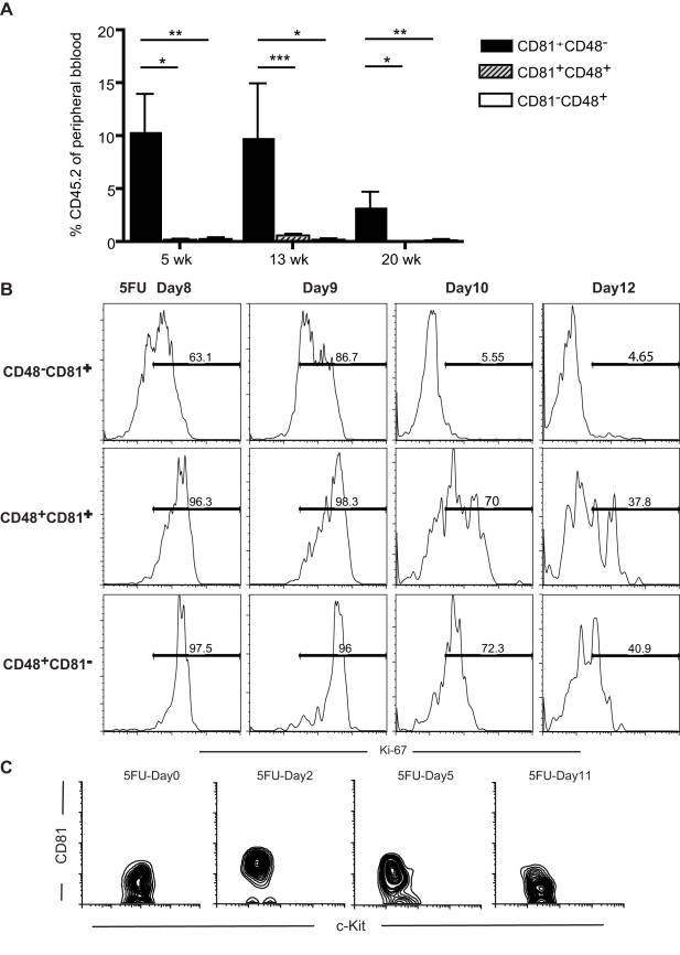
Figure 1. CD81 marks regenerating HSCs that are returning to quiescence.
(A) Competitive transplantation assay shows that among the SP cells from the 5FU-treated mice, the CD81+CD48− fraction contains most of the stem cell activity, compared with that of the CD81+CD48+ and CD81−CD48+ fractions. In this assay, 300Lin−SP (Side Population) cells expressing the indicated markers were transplanted with 2.5 × 10⁵ whole bone marrow competitors. The test donor population was derived from CD57Bl/6-CD45.2 mice, while the competitors and recipients were C57BL/6-CD45.1 mice, enabling us to determine the contribution of HSCs to peripheral blood production, using flow cytometry to detect the CD45.2 marker. The values are means ± SD (n = 5 per group per time point), **p<0.01, *p<0.05. (B) CD81+CD48−Lin−SP cells show the least expression of Ki-67, a proliferation antigen, at each 5FU time point tested, indicating that CD81+CD48− cells are the cells returning to quiescence most rapidly among the Lin−SP cells. For each time point, femurs and tibias from 3 to 4 mice were collected in each experiment, and at least two independent experiments were performed for each time point. (C) Expression of CD81 by HSCs (SP cKit+Lin−Sca1+, SPKLS) is upregulated when the stem cells are proliferating in response to 5FU stimulation (shown here are 5FU-Day2 and 5FU-Day5). CD81 is expressed at background levels in quiescent HSCs (5FU-Day0 and 5FUDay11), but is upregulated during proliferation (starting at 5FU-Day2).

