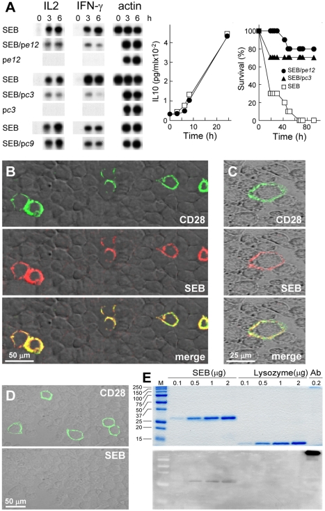
Figure 2. SEB binds to CD28.
(A) Phage display peptides selected by affinity for the SEB binding site in CD28 are SEB antagonists that protect mice from killing by SEB. PBMC were induced with SEB alone or with 0.1 µg/ml pc3, pe12, or pc9, a negative control. IL2 and IFN-γ mRNA are shown; β-actin mRNA indicates equal loading of RNA. For pe12, IL10 was determined (data are shown as means ± SEM (n = 3 experiments)). Mice (n = 10 per group) were challenged with 6 µg SEB alone or with 0.2 µg pe12 or 0.5 µg pc3; p for survival, 10−4. (B–D) Binding of SEB to cell surface CD28. Representative fields of confocal microscopy are shown. In (B), HEK293-T cells were transfected to express CD28-GFP fusion protein (green) and after 48 h incubated for 1 h with Alexa-Fluor-633-labeled SEB (red). In (C), BHK-21 hamster cells were transfected with CD28 cDNA vector and after 48 h incubated successively for 30 min with labeled SEB (red), goat polyclonal αCD28, and Cy2-labeled donkey anti-goat IgG (green). In (D), BHK-21 cells were transfected to express CD28 and after 48 h incubated first with goat polyclonal αCD28 and Cy2-labeled donkey anti-goat IgG (top) and only then with labeled SEB (bottom). (E) Binding of SEB to CD28. SEB, lysozyme, and polyclonal αCD28 (Ab) were separated on duplicate SDS-PAGE gels. Coomassie blue staining (top); far-western blot with CD28-Fc (bottom); M, size marker.

