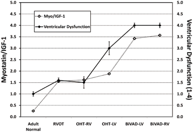
Figure 3. Myostatin to IGF-1 Ratio.
The relationship of myostatin/IGF-1 (left axis) and ventricular dysfunction (right axis) has been plotted for Adult Normal (n = 5), pediatric right ventricular outflow tract (RVOT) (n = 3), pediatric orthotopic heart transplant (OHT) (n = 7 paired LV and RV), and pediatric biventricular assist device (BiVAD) myocardial tissue samples (n = 3 paired LV and RV). A strong association between increased myostatin/IGF-1 ratios and worsening ventricular dysfunction exists: Adult Normal samples had low myostatin/IGF-1 levels, while BiVAD samples displayed high myostatin/IGF-1 levels.

