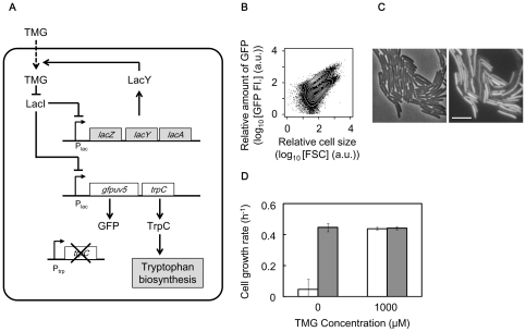
Figure 1. Rewired E. coli cell.
A. Modified lactose utilisation network with the rewired trpC. Influx of TMG is mediated by lactose permease LacY. Lactose repressor LacI inhibits lactose promoter Plac and is inactivated by TMG. gfpuv5 and trpC coding green fluorescent protein and phosphoribosylanthranilate isomerase, respectively, under the control of Plac. B. Scatter plot merged with contours of the cell population in the presence of tryptophan with 300 µM TMG obtained by flow cytometry. GFP FI and FSC (forward scattering) represent the fluorescence of green emission from GFP and relative cell size, respectively. C. Microscopic images of cell populations indicated in B. The phase-contrast and fluorescent images are shown on the left and right, respectively. Scale bar, 5 µm. D. Growth rate of the induced or suppressed cells in the presence (closed bars) or absence (open bars) of tryptophan. The suppressed and induced cells were prepared in the absence of TMG and by induction with 1,000 µM TMG, respectively. Error bars represent standard deviation for 3 replicates.

