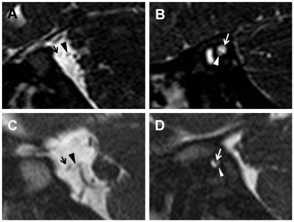
Figure 3. The parasagittal images of temporal MRI in patients with CHARGE syndrome.
(A–B) The cochleovestibular nerve (arrowheads) is larger in diameter than the facial nerve (arrows) at the cerebellopontine angle (A) and within the internal auditory canal (B) in Patient 1. (C–D) The cochleovestibular nerve (arrowheads) is smaller in diameter than the facial nerve (arrows) at the cerebellopontine angle (C) and within the internal auditory canal (D) in Patient 4.

