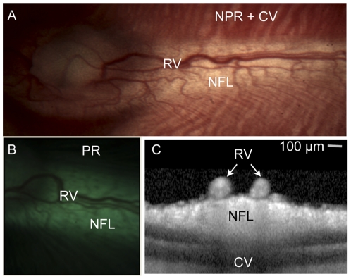
Figure 1. Illustration of rabbit retinal anatomy in one representative albino (panel a) and pigmented (panel b) animal.
(A,B) Color photographs of the retina at low magnification illustrate the relationship between the retinal vessels (RV), non-pigmented nerve fiber layer (NFL), non-pigmented retina (NPR) with visible choroidal vessels (CV) and pigmented retina that obscures underlying choroidal vessels (PR). (C) Optical coherence tomography of the retina in cross-section demonstrates that the retinal vessels lay on top of the unpigmented, myelinated nerve fiber layer. Note the complete lack of pigmentation in the albino animal versus the pigmented animal. Scale bar 100 microns (for panel C only).

