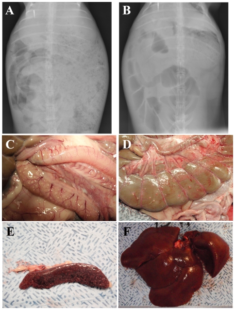
Figure 6. Gastrointestinal pathology and hepatosplenomegaly associated with tularemia in rabbits.
Anterioposterior radiographs showing the abdomen of a single representative rabbit two days prior (A) and four days after (B) infection with F. tularensis, illustrating gas production and distension of the intestines and stomach. Pictures of the small intestines (C), colon (D), spleen (E) and liver (F) examined at necropsy from a single representative rabbit.

