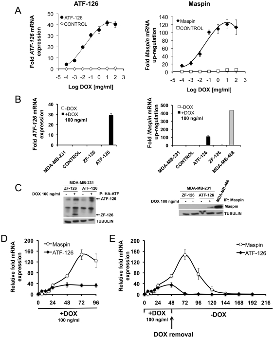
Figure 1. Induction of ATF-126 by DOX results in reactivation of the target gene Maspin.
A. Dose-response plot monitoring ATF-126 (left panel) and Maspin (right panel) mRNA levels upon treatment with increasing concentrations of DOX. CONTROL and ATF-126 cells were treated for 72 hours and mRNA was measured by quantitative real-time PCR (qRT-PCR). B–C. ATF-126 and Maspin mRNA expression levels by qRT-PCR (B) and western blot (C) induced with 100 ng/ml of DOX. MDA-MB-468 is a poorly aggressive ER- breast cancer cell line expressing endogenous Maspin as a reference control [12]. D. Time course kinetics of ATF-126 and Maspin mRNA levels by qRT-PCR upon DOX treatment. ATF-126 cells were induced with DOX and collected at 0, 6, 12, 18, 24, 48, 72 and 96 hours. E. Time course kinetics of ATF-126 and Maspin expression levels by qRT-PCR upon DOX treatment and removal. ATF-126 cells were induced with DOX for 48 hours, then DOX was removed from the media and cells were maintained in DOX-free media for an additional 168 hours. Gene expression levels were normalized to the −DOX cells. Data represents the mean ± SD of three independent biological replicates. MDA-MB-231-LUC are un-transduced cells; CONTROL, cells transduced with an empty vector; ATF-126, a full length ATF containing the 6 ZF DNA-binding domains and VP64 activator domain; ZF-126, a truncated or inactive ATF-126 lacking the VP64 activator domain.

