FIGURE 5:
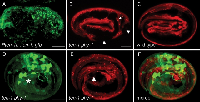
Epidermal defects correlate with defects in body wall muscles in arrested ten-1 phy-1 embryos. (A) TEN-1 is expressed in dorsal epidermal cells during embryogenesis. (B, C) Confocal images of embryos expressing mCherry under the muscle-specific myo-3 promoter. (B) Double-mutant embryo arrested at the 2.5-fold stage. Muscle strands are disrupted (arrowhead) and detached from the epidermal cells (arrow). (C) Wild-type embryos at the threefold stage show continuous strands of body wall muscles from head to tail. (D–F) Three-dimensional representation combining the epidermal (green) and muscle (red) markers in an arrested ten-1 phy-1 embryo. (D) Expression of pten-1b::gfp. The major epidermal defect is indicated by an asterisk. (E) Expression of pmyo-3::mCherry. The disrupted muscle strand is indicated by an arrowhead. (F) Merged image showing that the epidermal and muscle defects are in close proximity. Scale bar, 10 μm.

