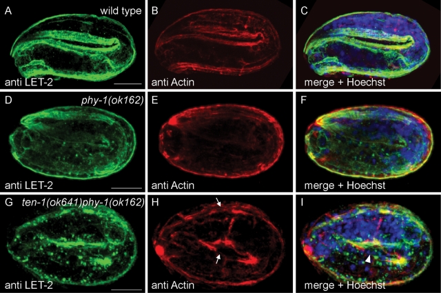
FIGURE 7:
Loss of phy-1 function causes aggregation of collagen IV in body wall muscles. Immunostaining with antibodies against LET-2 (green) and actin (red). DNA is stained with Hoechst dye (blue). (B, E, H) Longitudinal actin bundles correspond to the position of body wall muscles. (A, C) LET-2 is almost completely secreted into the BM in wild-type embryos at the threefold stage. The focus is on the BM separating muscles from epidermal cells. No aggregates of LET-2 can be detected in muscles. (D, F) phy-1(ok162) single mutants at the threefold stage frequently show intracellular aggregates of LET-2 as spots in close proximity to muscle nuclei. (G, I) In arrested ten-1 phy-1 double-mutant embryos, cluster formation of LET-2 protein is enhanced. (H) Actin bundles appear disorganized and are disrupted in some places (arrow). Similar results were obtained with anti EMB-9 antibody and are represented in Supplemental Figure S4. Scale bar, 10 μm.

