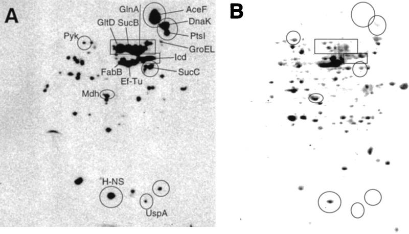
Figure 5.
Specific protein carbonylation determined by two-dimensional Western blot immunoassay. (A) An autoradiograph obtained after carbonyl immunoassay of proteins from a wild-type E. coli culture starved for 1 day; (B) the same protein extract blotted to PVDF membrane and stained with Coomassie brilliant blue. (GltD) Glutamate synthase; (GlnA) glutamine synthetase; (Icd) isocitrate dehydrogenase; (SucB) dihydrolipoamide succinyltransferase; (Mdh) malate dehydrogenase; (AceF) dihydrolipoamide acetyltransferase; (SucC) succinyl CoA ligase; (PtsI) phosphoenolpyruvate–protein phosphotransferase; (Pyk) pyruvate kinase; (UspA) universal stress protein A; (FabB) β-ketoacyl-[acyl carrier protein] synthetase; (EF-Tu) elongation factor Tu. The analysis was repeated two times to confirm reproducibility. Representative results are presented.

