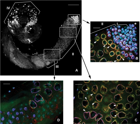
FIGURE 1:
Nuage granules of at least two types are detected in the perinuclear area of primary spermatocytes. Spatiotemporal pattern of nuage and piNG-bodies in the testes. (A) A full-size yw testes. White boxes indicate positions of the fragments enlarged in B–D. Scale bar, 100 μm. Brackets in A and B indicate different germinal cells: I, spermatogonial cells; II, spermatocytes; III, round spermatids; IV, elongated spermatids. Asterisk indicates the germinal proliferative center. Testes of yw flies were stained with anti-Vasa (green), anti-Aub (red), and anti-lamin (violet) antibodies; chromatin was stained with DAPI (blue). Colocalization of green (Vasa) and red (Aub) signals yields yellow color. (B) Testis apical tip. Small nuage granules (<1 μm) form discontinuous rings around the nuclei. Large nuage granules, the piNG-bodies (2.38 ± 0.35 μm), are indicated with white arrowheads. Note that not all large nuage granules can be seen on a single confocal slice. Nuage first appears in germinal stem cells and spermatogonial cells, whereas piNG-bodies are formed later in primary spermatocytes at the S2b stage (nuclear diameter of ∼6–10 μm). See also Supplemental Figure S1A for separate channel presentation. (C) The S5 stage (nuclear diameter of ∼16–20 μm). The piNG-bodies are indicated with white arrowheads. See also Supplemental Figure S1B for separate channel presentation. (D) Spermatocytes at the end of the S stage (nuclear diameter of ∼15 μm) and round spermatids (nuclear diameter of ∼4–6 μm). Nuage is preserved at the end of the S stage; however, prominent piNG-bodies are absent. No detectable nuage structure can be seen in round spermatid cells. See also Supplemental Figure S2 for separate channel presentation. Scale bars for B–D, 15 μm.

