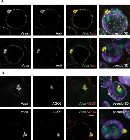
FIGURE 3:
Mutual arrangement of Vasa, Aub, and AGO3 within the piNG-body. (A) A high level of Vasa (green) and Aub (red) colocalization in the piNG-bodies. Two different piNG-bodies stained with Vasa (green) and Aub (red) as well as the corresponding pseudo–three-dimensional (3D) images are shown. Note that both Vasa and Aub occupy the periphery of the piNG-body rather than the internal space. (B) AGO3 protein is found in the piNG-body core and colocalizes with Vasa only along the boundary line regions. Two different piNG-bodies stained with Vasa (green) and AGO3 (red) and the corresponding pseudo-3D images are shown. Testes of yw flies were stained with anti-Vasa (green), anti-Aub (A), or anti-AGO3 (B) (red) and anti-lamin (violet) antibodies; chromatin was stained with DAPI (blue). Colocalization of green and red signals yields yellow color. Scale bars, 3 μm. For more images of AGO3 and Vasa distribution in the piNG-body in the isosurface format see Supplemental Figure S5.

