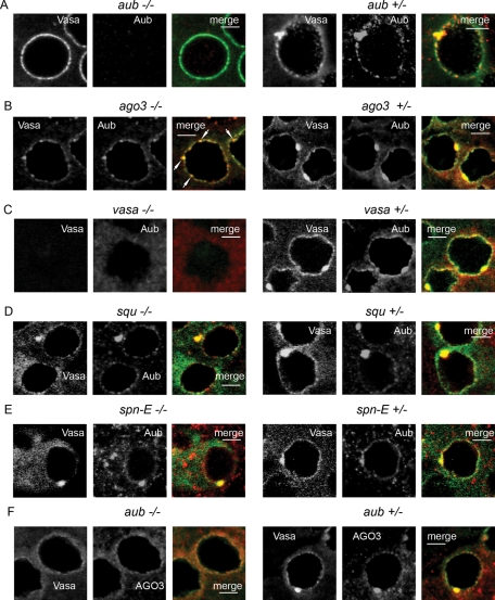
FIGURE 6:
Mutational analysis of the piNG-body. In all experiments, the testes were stained with anti-Vasa (green) and anti-Aub (red) antibodies, except for F, in which they were stained with anti-AGO3 (red) antibodies. Scale bars, 5 μm. (A, F) The aub mutation effect on the piNG-body formation. In the aubHN/aubQC42 testes, small AGO3- and Vasa-stained nuage granules are visible, but no piNG-body formation is observed. (B) The ago3 mutation effect on the piNG-body formation. No normal-sized piNG-bodies are visible; however, up to several Aub/Vasa–stained particles of ∼1 μm size are detected on the nuclear surface or in the cytoplasm of spermatocytes (white arrows). (C) No nuage is found in the testes of vasEP812/vasD1 flies (the same results were obtained with vasEP812/vasPH165 and vasEP812 mutants). (D) piNG-bodies and conventional nuage granules are distinctly seen in the squPP32/DfExel7066 transheterozygous flies. (E) piNG-bodies and conventional nuage granules are preserved in the spn-E616 homozygous flies.

