
Figure 8.
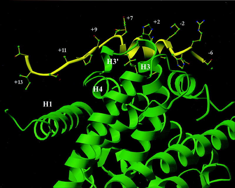
Model of SRC-1 LXD2 (−6) through (+14) bound to BRL49653-liganded PPARγ LBD based on the co-crystal structure with SRC-1 amino acids 623–710 (Nolte et al. 1998). A ribbon drawing of the LXD2 motif of human SRC-1 is shown in yellow with the human PPARγ LBD shown in green. When the electron density maps of the co-crystal structure of the SRC-1 heterodimer with liganded PPARγ (Nolte et al. 1998) are examined and modeled, the +12, +13 amino acids form weak interactions at the amino terminus of helix 1; the +6 side chains contact the carboxyl terminus of helix 3; amino acids +9 and +10 form interactions with the small helix (H3′) between helices 3 and 4.