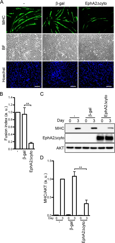
FIGURE 7:
IGF-I–induced myogenic differentiation is blunted by blocking the ephrinA/EphA signal. (A) C2C12 myoblasts infected without (−) or with adenoviruses encoding either β-gal or EphA2Δcyto were differentiated into myotubes in DMEM containing 1% FBS and 10 nM IGF-I for 3 d. The cells were immunostained with anti-MHC antibody and visualized with Alexa Fluor 488–conjugated secondary antibody. The cells were also poststained with Hoechst 33342 to visualize the nuclei. Alexa 488 (MHC), bright-field (BF), and Hoechst 33342 (Hoechst) images are shown as indicated at the left. Experiments were repeated three times with similar results. Scale bars, 100 μm. (B) The fusion index observed in A was determined as described in the legend of Figure 3B. Values are expressed relative to that observed in the uninfected cells and shown as means ± SD of three independent experiments. (C) Adenovirus-infected C2C12 myoblasts were differentiated into myotubes as described in A for time periods (days) indicated at the top. Cell lysates were subjected to Western blot analysis using anti-MHC (MHC), anti-HA (EphA2Δcyto), and anti-AKT (AKT) antibodies as indicated at the left. (D) The expression level of MHC observed in C was quantified by normalizing the expression of MHC by that of AKT. Values are expressed relative to that observed in the uninfected cells differentiated for 3 d and shown as means ± SD of three independent experiments. **p < 0.01, significant differences between two groups. a.u., arbitrary unit(s).

