
Figure 3.
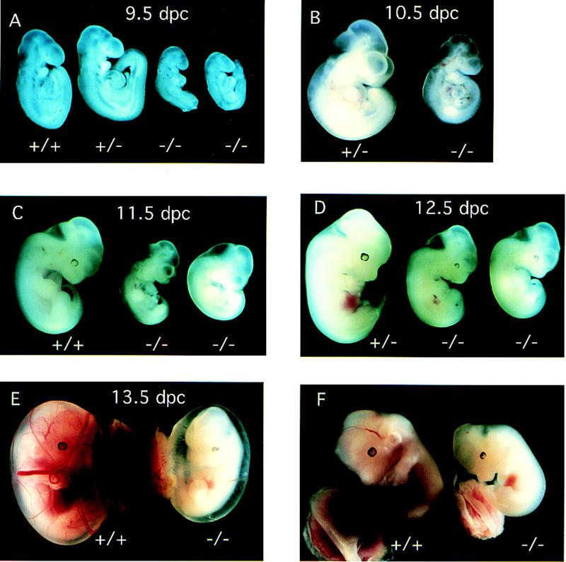
Blm−/− embryos exhibit small size and developmental delay. (A) 9.5 dpc; a pair of normal (Blm+/+ and Blm+/−) embryos are compared with two Blm−/− embryo littermates. (B) 10.5 dpc; comparison of Blm+/− and Blm−/− embryo littermates. (C,D) Normal 11.5-dpc Blm+/+ and 12.5-dpc Blm+/− embryos are compared with pairs of mutant Blm−/− littermates. (E) Comparison of 13.5-dpc Blm+/+ embryos with a mutant Blm−/− littermate with intact yolk sac and placenta. (F) The same pair of embryos shown in E with yolk sacs removed. Embryos in A were photographed in 70% ethanol after fixation in 4% paraformaldehyde in PBS. Embryos in (B–F) were photographed at the time of dissection. At dissection, embryos were determined to be alive on the basis of the presence of a heartbeat.