
Figure 5.
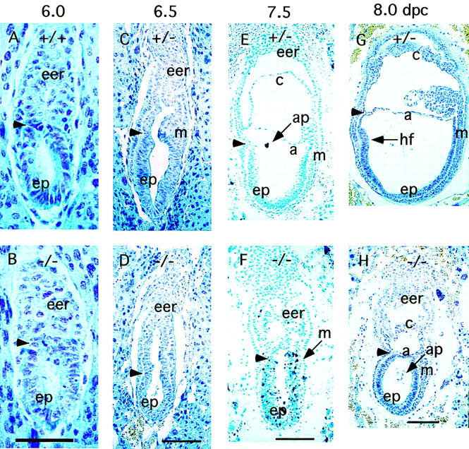
Defective development in Blm mutant embryos. Histological analysis was performed on embryo sections stained with Feulgen and fast green (A–D,G,H) or methyl green (E,F). Each pair of panels represent embryos derived from the same litter; developmental stage is indicated (top). (A,B) 6.0-dpc early egg cylinder stage of Blm+/+ and Blm−/− embryos, respectively. (C,D) 6.5-dpc gastrulation stage of Blm+/− and Blm−/− embryos, respectively. (E) TUNEL assay of a 7.5-dpc Blm+/− embryo; apoptotic debris is indicated (ap, arrow). (F) TUNEL assay of a 7.5-dpc Blm−/− embryo; black apoptotic nuclei appear throughout the epiblast. (G,H) Early organogenesis stage of 8.0-dpc Blm+/− and Blm−/− embryos, respectively. Apoptotic debris is observed in the amniotic cavity of the mutant [(H) ap, arrow]. (Arrowheads) Separation between the extraembryonic and embryonic region. (a) amnion; (c) chorion; (eer) extraembryonic region; (ep) epiblast; (hf) headfold; (m) mesoderm. Size bar, 60 μm in A and B; 120 μm in C–F; 250 μm in G and F.