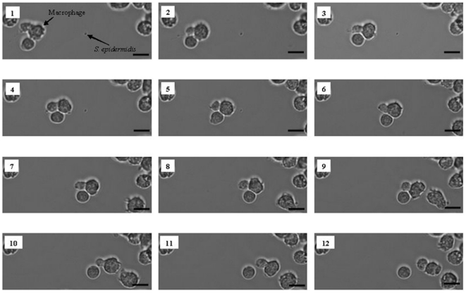
Figure 3. Macrophage migration towards S. epidermidis and phagocytosis under flow.
Phase-contrast images of macrophage activity toward S. epidermidis ATCC 35983 on a PMMA surface in the presence of U2OS cells: macrophage migration towards S. epidermidis (images 1–5), bacterial clearance by phagocytosis (images 6–7) and further migration (images 8–12). The bar denotes 50 µm.

