
Figure 5.
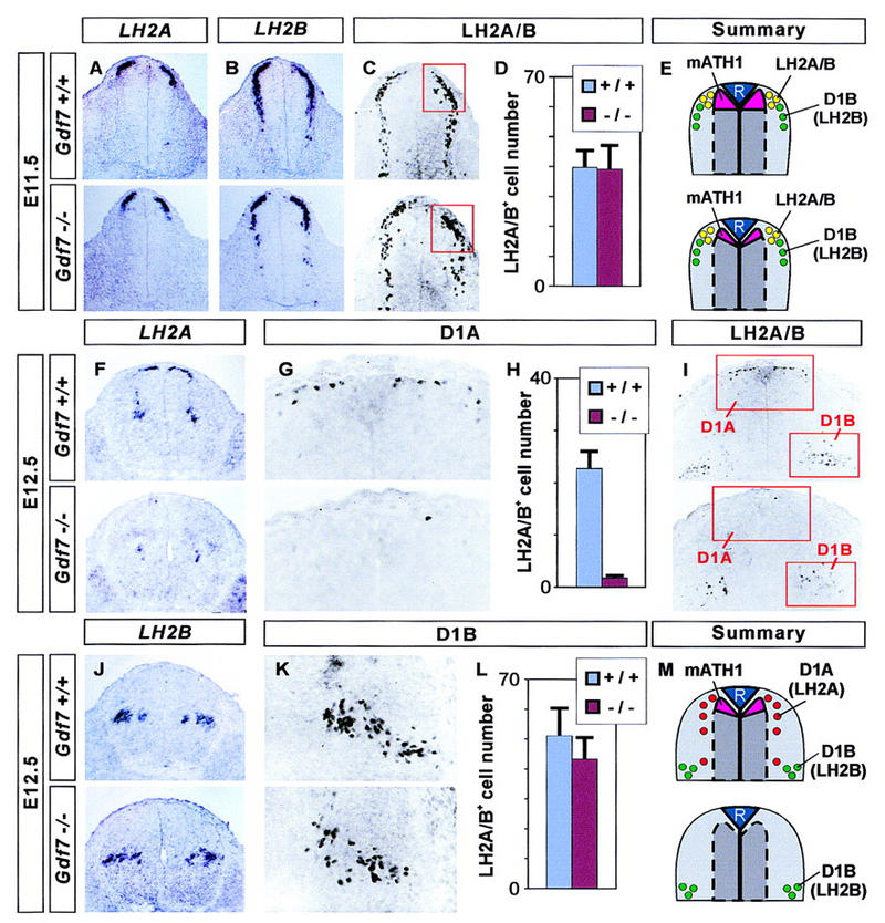
A selective loss of D1A neurons in Gdf7 mutant mice. Analysis of dorsal neuron populations in the cervical (forelimb level) spinal cord in wild-type embryos (top panels) and in Gdf7m1 homozygous mutant embryos (bottom panels). (A–E) D1 neuron populations in the E11.5 spinal cord (summarized in E). In situ hybridization shows that at this stage, expression of LH2A (A) and LH2B (B) is normal in Gdf7 mutant embryos. The number of dorsal LH2A/LH2B-immunoreactive neurons (red box in C) is not changed in Gdf7 mutants. (D) Quantitative analysis of the dorsal LH2A+/LH2B+ cell population (mean ± s.d., n = 10–25 sections from two to six embryos of each genotype). We also found no alteration in the number of more ventrally located D1B neurons in Gdf7m1 mutants. (F–M) D1 neuron populations at E12.5 (summarized in M). At this stage, expression of LH2A (F) is greatly reduced, whereas LH2B (J) is unaffected in Gdf7 mutant embryos. Similarly, the number of dorsal LH2A/LH2B-immunoreactive cells (D1A neurons; boxed in I, shown enlarged in G) is reduced to ∼8% the number in wild-type and the number of ventral LH2A/LH2B-immunoreactive cells (mostly D1B neurons; boxed in I, enlarged in K) is not significantly altered in Gdf7 mutants. (H,L) Quantitative analysis of the D1A and D1B cell populations (mean ± s.d., n = 10–25 sections from two to six embryos of each genotype).