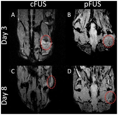
Figure 2. Evaluation of FL-SPION-labeled macrophage migration to FUS-treated muscle tissue by MRI.
T 2*-weighted MR images were obtained at 3T after 3 (A and B) and 8 (C and D) days post-FUS. Imaging reveals hypointense voxels in regions of the right leg that were treated with cFUS (A and C) or pFUS (B and D) exposures.

