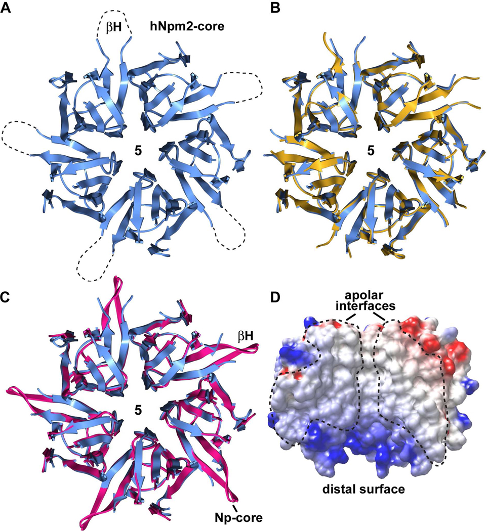
Figure 2. Npm2- and Np-core pentamers.
(A) The Npm2-core pentamer is shown as a ribbon representation viewed from the top. Approximate positions of β-hairpin tips are indicated with dotted loops, since they are not well ordered. (B) Two independent Npm2-core pentamers in the asymmetric unit are over-layed. (C) A perfect Np-core pentamer with 5 β-hairpins (in red) 9 is shown over-layed with the human Npm2-core pentamer from panel A (in blue). (D) An electrostatic surface is shown of three adjacent subunits within the Npm2-core pentamer. The exposed subunit-subunit interface is non polar (see dashed outlines).

