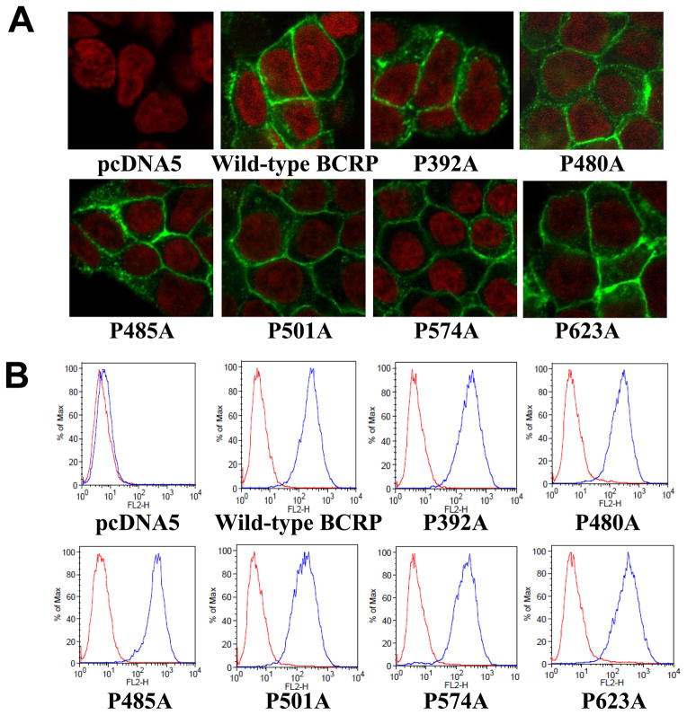
Figure 3. Confocal microscopy analysis and cell surface expression of Flp-In™-293 cells stably expressing wild-type and mutant BCRP.
A) The cellular localization of wild-type and mutant BCRP in Flp-In™-293 cells (shown in green) was determined by immunofluorescent confocal microscopy using the BCRP-specific mAb BXP-21. Cell nuclei were stained with DAPI and are shown in red. B) Expression of wild-type and mutant BCRP on cell surface of stably transfected Flp-In™-293 cells was detected using the 5D3 monoclonal antibody. Representative flow cytometry histograms showing cell surface expression of wild-type and mutant BCRP are presented. The red and blue peaks represent the phycoerythrin fluorescence associated with cells treated with the IgG2b negative control and the 5D3 antibodies, respectively. No surface expression of BCRP was detected in the pcDNA5 vector control cells.

