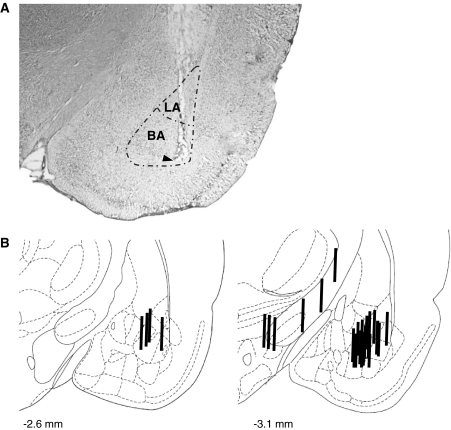
Fig. 1.
Probe placements in the amygdala. a A representative photomicrograph of a cresyl-stained coronal section showing the localization of the tip of the membrane of a microdialysis probe (arrowhead) within the BA/LA. b Schematic drawings of the localization of the microdialysis membranes (bold black lines) used in the present study within and outside of the BA/LA. BA basal amygdala, LA lateral amygdala. Illustrations modified from Paxinos and Watson (1998)

