Figure 5.
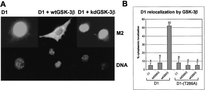
GSK-3β overexpression redirects cyclin D1 to the cytoplasm. (A) NIH-3T3 cells transiently expressing CDK4 and either Flag-tagged cyclin D1 (left), Flag-tagged cyclin D1 plus wild-type GSK-3β (wtGSK–3β; middle), or Flag-tagged cyclin D1 plus kinase-defective GSK-3β (kdGSK-3β; right) were fixed and processed for immunofluorescence. Flag-tagged cyclin D1 was visualized by staining with the M2 monoclonal antibody to the tag (top), and cellular DNA was stained with Hoechst dye (bottom). (B) The subcellular localization of Flag-tagged cyclin D1 or Flag-tagged D1-(T286A) without ectopic GSK-3β or with either wtGSK-3β or kdGSK-3β was determined by immunofluorescent staining with the M2 monoclonal antibody as above. The percentage of cells expressing exclusively cytoplasmic cyclin D1 or D1-(T286A) is presented graphically and represents the average of at least four independent experiments. Vertical bars indicate standard deviations from the mean.

