
Figure 4.
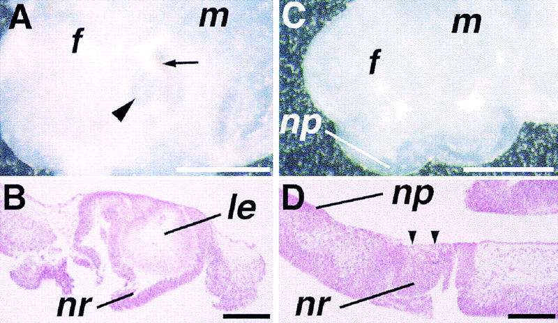
Morphology of eye explants in vitro. (A) A wild-type eye primordium taken at the 20-somite stage and cultured for 4 days. Not only the lens (arrowhead) but also surrounding retina and pigmented epithelium (arrow) are easily recognized under the dissection microscope. (B) Histological section of the eye explant shown in A. A well-differentiated lens (le) is seen, surrounded by the neuroretina (nr) of the optic cup. (C) An eye primordium from a 9.5-dpc mutant embryo cultured for 5 days. No lens formation is recognized macroscopically. (D) Histological section reveals that the lens is absent in the mutant eye primordium at the site of contact between the ectoderm and prospective neuroretina (arrowheads), whereas the nasal placode (np) is developed in the adjacent ectoderm. Bar, 500 μm (A,C); 100 μm (B,D).