
Figure 1. a & b. [GR expression in control and FBRKO mice].
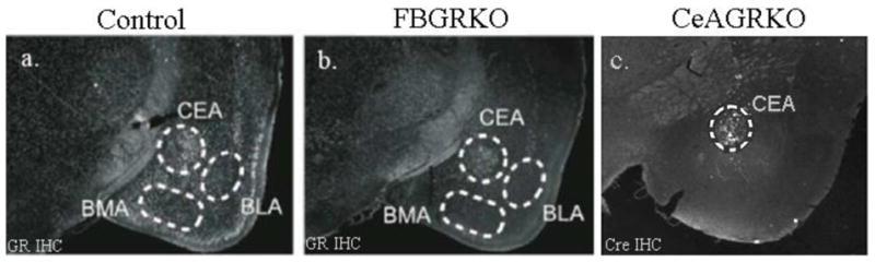
GR immuno-reactivity (white nuclei) is detected throughout neuronal populations in control mice (a.), but is markedly reduced or absent in the cortex, basolateral nucleus of amygdala (BLA) and basomedial amygdala (BMA) in FBGRKO mice. Note that GR expression is intact in the CeA and thalamus of the FBGRKO mice. c. [Cre-recombinase and GR staining in CeAGRKO mice]. Immunohistochemistry showing Cre immunoreactivity 2 weeks after LV-Cre infection