
Figure 4.


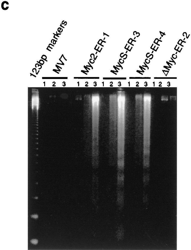
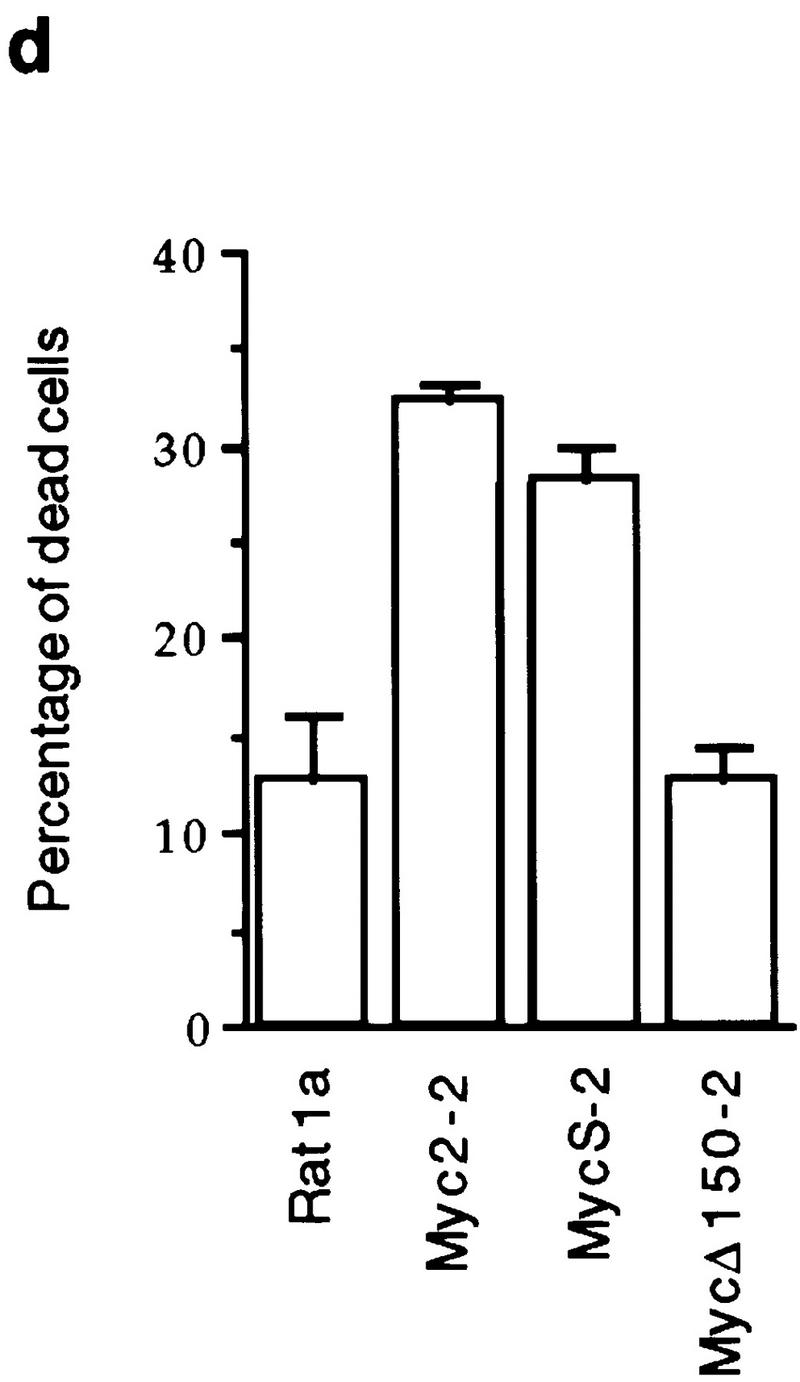
c-MycS induces apoptosis. (a) Apoptosis of Rat 1a cells induced by c-Myc–ER proteins. The stable Rat 1a cell lines expressing c-Myc2–ER, c-MycS–ER, c-MycΔ106–143 (ΔMyc–ER), or the pMV7 vector alone were seeded at 5 × 10⁴ cells in replicate 35-mm plates. After 24 hr, the cells were transferred into DMEM containing 10% CS with or without 1 μm OHT. At the indicated time points, triplicate dishes were counted. The number of floating cells was divided by the total cell count to yield the percentage of dead cells. (b) Apoptosis induced by c-Myc–ER proteins in low serum. The cell lines described above were seeded at 5 × 10⁴ cells in replicate 35-mm plates. After 24 hr, the cells were transferred into DMEM containing 0.25% CS with or without 1 μm OHT. At the indicated time points, the number of dead cells was determined as described above. (c) DNA fragmentation analysis of c-Myc–ER-expressing cells. The cell lines described above were plated at 1.5 × 10⁶ cells/100-mm dish in DMEM containing 10% FCS. After 24 hr, the medium was changed to DMEM containing 0.25% FCS with or without 1 μm OHT. After 20 hr, the adherent cells were combined with the nonadherent cells and the DNA was extracted and analyzed by agarose gel electrophoresis as described. (Lanes 1) Cells in DMEM plus 10% FCS; (lanes 2) cells in DMEM plus 0.25% FCS without OHT; (lanes 3) cells in DMEM plus 0.25% FCS with OHT. (d) Apoptosis induced by constitutive c-Myc proteins in low serum. Rat 1a cells or Rat 1a stable cell lines expressing either c-Myc2, c-MycS or c-MycΔ150 were seeded at 5 × 10⁴ cells in replicate 35-mm plates. After 24 hr, the cells were transferred into DMEM without serum, and the number of dead cells was determined as described above.