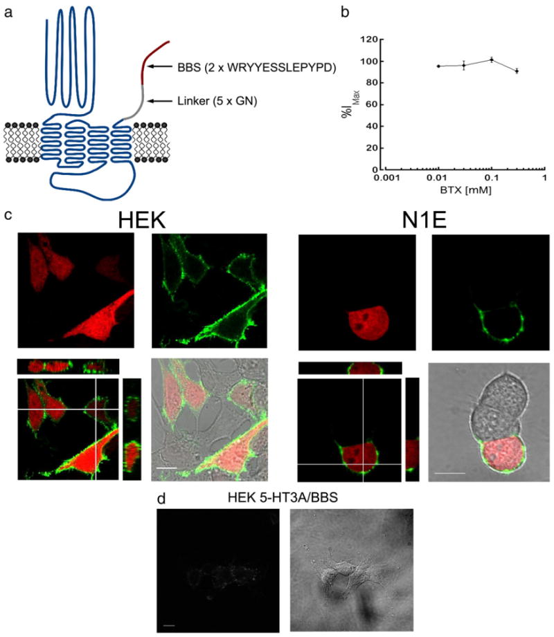
Fig. 1.

α-bungarotoxin conjugated to Alexa-fluorophores specifically labels surface 5-HT3A/BBS receptors. a) Schematic representation of the 5-HT3A/BBS receptor. b) Whole-cell voltage clamp configuration was obtained with HEK-293 cells stably expressing 5-HT3A/BBS receptors. 5-HT3A/BBS mediated currents were recorded in the absence or presence of varying concentrations of non-conjugated BTX. The peak amplitudes of the currents in the presence of BTX are expressed as a percent of the peak amplitudes in the absence of BTX. c) HEK-293 or N1E-115 cells transiently transfected with 5-HT3A/BBS and pmCherry. Transfected cells identified by the expression of mCherry (red) and 5-HTA/BBS receptors are labeled with BTX/488 (green). Merged images include XY, XZ, and YZ images to show that BTX/488 labeling is consistently at or near the cell surface with intracellularly expressed mCherry. d) HEK-293 cells stably expressing 5-HT3A/BBS receptors incubated in non-conjugated BTX for 30 min at 4 °C prior to incubation with Alexa/488 conjugated BTX for 30 min at 4 °C. Scale bars=10 μm.
