
Fig. 7.
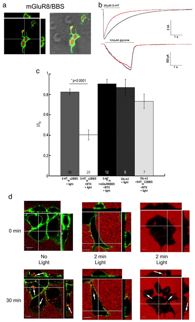
Collateral damage of receptor internalization but not receptor function. HEK-293 cells were transiently transfected with wild-type 5-HT3A, metabotropic glutamate 8 receptors (mGluR8) tagged with the BBS and pmCherry. a) Surface mGluR8 receptors were labeled with BTX/488 and cells were fixed with 4% formaldehyde then imaged. b) Representative traces of currents mediated by 5-HT3A receptors (top traces) and glycine α2 receptors (bottom traces) before (black traces) and after (red traces) BTX/488 labeled mGluR8/BBS or 5-HT3A/BBS receptors respectively were exposed to 9.73 mW/cm2 of ∼488 nm light. c) Averaged cumulative peak amplitudes evoked by either 30 μM 5-HT or 100 μM glycine expressed as a proportion of the initial peak amplitude (I/I0) after 9.73 mW/cm2 of ∼488 nm light from 5-HT3A/BBS, 5-HT3A, or glycine receptors in the presence or absence of increased oxygen. d) Surface 5-HT3A/BBS receptors were labeled with BTX/488 and transferrin/555(red) was added to the medium (top row). Cells were maintained at 37 °C on the microscope and imaged after 30 min (bottom row). Cells with BTX/488 (green) labeled 5-HT3A/BBS receptors were not exposed to light for 2 min (left column) or exposed to ∼488 nm light every 5 s for 2 min (middle column). Cells without BTX/488 were exposed to ∼488 nm light every 5 s for 2 min (right column). Arrows indicate internalized puncta. Scale bar=10 μm.