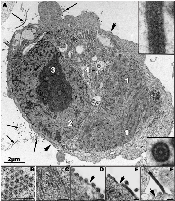
Figure 2.
Morphological characteristics of Zaire ebolavirus (ZEBOV) replication. (A) Ultrathin section of a ZEBOV-infected Vero cell containing large viral inclusions. The inclusions are composed of granular material (1; also shown in (C)) and rod-like nucleocapsids. Released virions are indicated by long arrows and are shown in the inserts. (B) Cross section of viral inclusion containing nucleocapsids. (C) Longitudinal section of viral inclusion containing nucleocapsids. (D–F) Budding of viral particles. In the initial step of budding a particle can be positioned parallel (D and E, cross and longitudinal sections), or perpendicular (F) to the membrane and then subsequently is enveloped. Short arrows indicate the cellular plasma membrane. 2—nucleus, 3—nucleolus, 4—Golgi zone. Bars in Figures 2B–F correspond to 250 nm. Thick part of frame around cross-sectioned virion corresponds to 120 nm, and thick part of frame around longitudinal section corresponds to 160 nm. Transmission electron microscopy. Cells were fixed at 16 h p.i.

