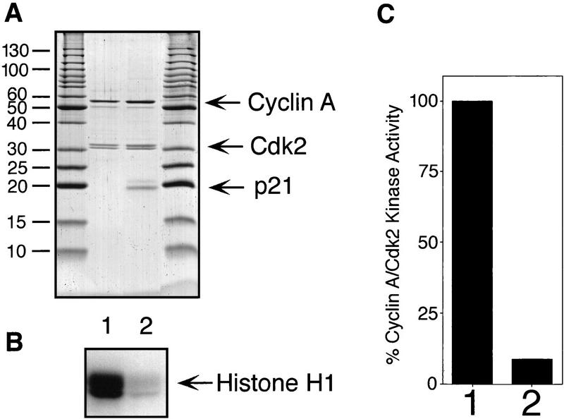
Figure 6.
p21/Cyclin A/Cdk2 complexes are kinase inactive. (A) Purified cyclin A/Cdk2 complexes (lane 1) and p21/cyclin A/Cdk2 complexes (lane 2) were separated by SDS-PAGE and stained with Coomassie brilliant blue to estimate relative concentrations of their polypeptides (A). Size markers are shown at the sides. The kinase activities of diluted samples (1:100) were determined using [γ-32P] ATP and histone H1 as substrates. The amount of 32P incorporation in the histone H1 bands is shown after SDS-PAGE by autoradiography (bottom) (B) and determined using a PhosphorImager (C). Kinase activities are shown as percentage of that of the noninhibited complexes and were normalized for the kinase subunits present in the complexes. (C).

