Fig. 2.
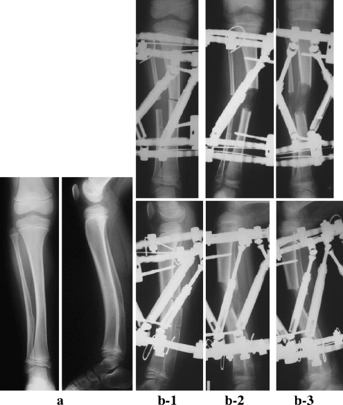
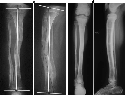
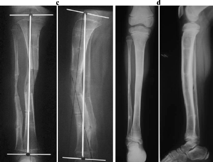
Case 2. a Radiographs before the operation at when the girl was aged 5 years. b 1 Radiographs just after the operation. b 2 Radiographs during deformity correction and lengthening (observe the worsened deformity). b 3 Radiographs after completion of the revised deformity correction and lengthening program. c Radiographs after removal of the Taylor spatial frame. d Radiographs at the final follow up


