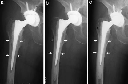
Fig. 2.
A 70-year-old woman with stem subsidence and decrease in the initial radiolucency. Initial radiolucency on the X-ray at 1 month after operation (a; white arrows) had already decreased at 6 months after operation (b) with stem subsidence. Stem subsidence of 2.52 mm had occurred by 1 year after operation (c), and radiolucency decreased more at zones 2, 3, 5, and 6. In addition, the border of cement and cancellous bone at zone 1 became indistinct (black arrow)

