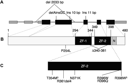
Figure 1.
GATA2 gene. (A) Genomic organization of GATA2 showing 2 5′-untranslated and 5 coding exons. Wider boxes represent coding regions. Insertion/deletion mutations predicted to result in null alleles are shown above. (B) Protein domains of GATA2, showing N- and C-terminal zinc fingers (ZF-1, ZF-2) and nuclear localization signal (N). (C) Missense and in-frame deletion mutations identified within ZF-2. Superscript numerals indicate the number of independent mutations.

