Figure 1.
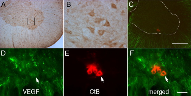
Representative photomicrographs of VEGF immunostaining in C4 phrenic motor neurons. A, DAB staining revealed VEGF expression in large, presumptive phrenic motor neurons (small black box) and interneurons. B, Higher magnification of small black box from A. C, CtB was injected intrapleurally to localize phrenic motor neurons (red cells in ventral horn). D–F, VEGF is expressed in phrenic motor neurons (note merged image of red CtB back-labeling and VEGF protein coexpression) but is also found in the surrounding neuropil. Sections incubated without primary or secondary antibodies served as negative controls. Scale bars: A, C, 400 μm; B, D–F, 50 μm.

