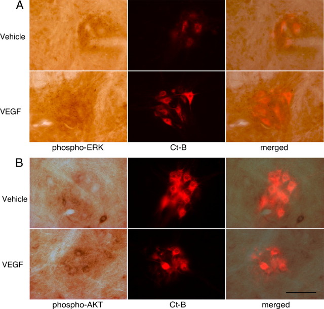
Figure 3.
Phospho-ERK and phospho-Akt expression in C4 phrenic motor neurons and upregulation after intrathecal VEGF injection. A, Phospho-ERK (far left panels, dark brown staining) is expressed in C4 phrenic motor neurons; staining tends to be punctate and bouton like. B, Phospho-Akt protein is colocalized with CtB-back-labeled phrenic motor neurons but appears to have a more dispersed, cytoplasmic distribution (red fluorescence). Both phospho-ERK and phospho-Akt staining increased after intrathecal VEGF injections (see Results). Sections incubated without primary or secondary antibodies served as negative controls. Data are means ± 1 SEM. *p < 0.05 versus vehicle. Scale bar, 100 μm.

