Figure 3.

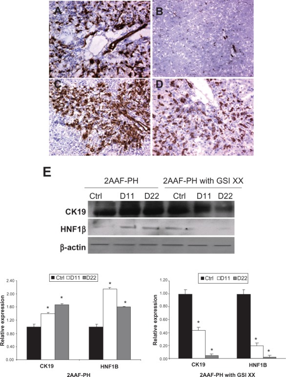
Analysis of oval cell surface marker expression upon Notch inhibition. A–D) Immunohistochemical analysis of OV-6 expression during 2AAF-PH alone (A and B) and in combination with GSI XX treatment (C and D). In the 2AAF-PH group, there is a dramatic increase in OV-6 expression at day 11 post-PH (A), which drops back down by day 22 post-PH (B), because all the oval cells have differentiated into mature phenotypes by this point. In the GSI XX-treated group similar levels of OV-6 expression are seen at day 11 post-PH (C), but a significant amount of staining remains at day 22 post-PH when Notch signaling is inhibited (D). E) Western blot analysis performed on protein isolated from liver taken at day 11 and day 22 post-PH from both treatment groups and probed with an antibody specific for the biliary markers CK19 and HNF-1β. During 2AAF-PH alone, expression of CK19 increases at day 11 post-PH and stays elevated by day 22 post-PH, indicative of biliary differentiation in the regenerated liver. However in the GSI XX-treated group, there is not as significant an increase in CK19 levels at day 11, a difference that is even more pronounced in the day 22 post-PH sample. Similar analysis performed with an antibody specific for HNF-1β shows a stark downregulation of the biliary transcription factor at both days 11 and 22 post-PH in the GSI XX-treated group as compared with 2AAF-PH alone. Bottom: Semiquantitative analysis of CK19 and HNF-1β protein in both treatment groups for control, day 11 post-PH, and day 22 post-PH samples. Expression was normalized to β-actin and significance calculated compared with control animals.
Notes: *P <0.01, error bars, SD.
Abbreviations: GSI XX, γ-secretase inhibitor; PH, partial hepatectomy; 2AAF-PH, 2-acetylaminofluorine implantation followed by 70% surgical resection of the liver; SD, standard deviation; HNF, hepatocyte nuclear factor.
