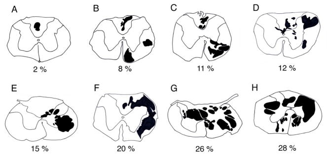
Figure 1.
Camera lucida drawings from C4 to C5 spinal segments of spinally contused rats at 21 days post-injury. Sections depicted had the largest cross-sectional area occupied by cyst-like cavitations (black objects). Injury size ranged from 2 to 28% (A to H) of total section area and most injuries had a unilateral bias.

