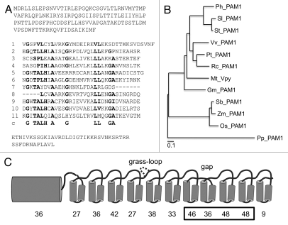
Figure 1.
Sequence analysis and phylogeny of VAPYRIN from diverse plants. (A) Predicted amino acid sequence of the petunia VAPYRIN protein PAM1. The 11 repeats of the ankyrin domain are aligned, and the ankyrin consensus sequence is shown below the eleventh ankyrin repeat (line c). Conserved residues that are characteristic for ankyrin repeats (Mosavi et al. 2004)17 are depicted in bold face. (B) Unrooted phylogenetic tree representing the VAPYRINs of eight dicot species (Petunia hybrida, Solanum lycopersicon, Solanum tuberosum, Vitis vinifera, Populus trichocarpa, Ricinus communis, Medicago truncatula and Glycine max) three monocot species (Sorghum bicolor, Zea mays and Oryza sativa), and the moss Physcomitrella patens. (C) Degree of conservation of the individual ankyrin repeats of VAPYRIN. Schematic representation of the MSP domain as N-terminal barrel-shaped structure, and of the individual ankyrin repeats as pairs of alpha-helices. An additional loop occurring only in monocots (grass-loop) is inserted above repeat 4, and the deletion between repeat 7 and 8 is indicated (gap). This latter feature is common to all VAPYRIN proteins. The percentage of amino acid residues that are identical in at least 11 of the 12 VAPYRINS is given below the MSP domain and the eleven ankyrin repeats. The box highlights repeats 7–10 which contribute to the predicted binding site (compare with Figs. 3 and 4).

