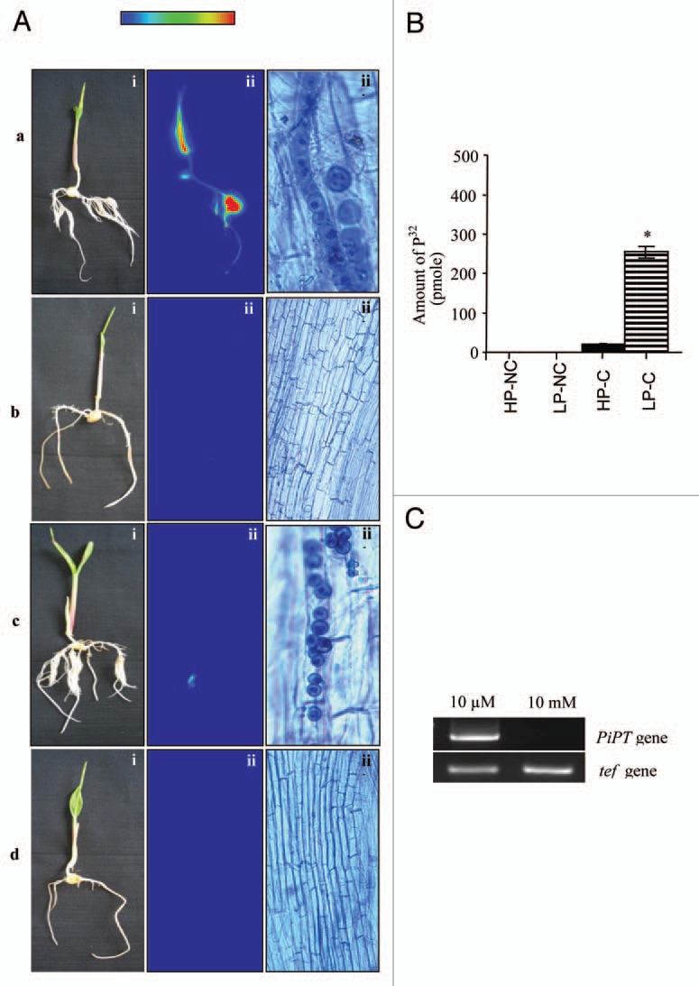
Figure 1.

Transport of P to maize plants by P. indica was carried out in bi-compartment Petri dish culture system.10 (A) Radioactivity incorporated in plant was demonstrated by autoradiography. Radioactivity counts intensities are shown in false color code (vertical bar, low to high). (i) Whole maize plant before autoradiography. (ii) False-color autoradiograph of the maize plant obtained after 3 h of exposure of the maize plant. (iii) Microscopic view of a sample of plant root before autoradiography. (a) Maize plant colonized with P. indica grown at 10 µM P concentration referred as P-deprived condition (LP-C). (b) Non-colonized maize plant grown at 10 µM P concentration, (c) Maize plant colonized with P. indica grown at 10 mM P concentration referred as P-rich condition (HP-C). (d) Non-colonized maize plant grown at 10 mM P concentration. Transport assay was done at 100 mM with 1 mM of P32 (specific activity 200 mCi/mmol). (B) Amount of 32P transferred into the maize plant components by P. indica. Bar labeled with the (*) represents significance as compared with the (HP-C) (p < 0.05). (C) PiPT gene expression analysis during colonization at P-deprived and P-rich condition. RT-PCR analysis was performed to check the PiPT gene expression in P. indica grown at P-deprived and P-rich condition. Lane 1 shows amplified PiPT DNA fragment (1.6 kb) from P. indica grown at P-deprived condition (10 mM). Lane 2, no expression of PiPT was observed when P. indica was grown at P-rich condition (10 mM). tef gene (220 bp) of P. indica was used as a loading control. A color version of this image is available at www.landesbioscience.com/journals/psb/article/15106/.
