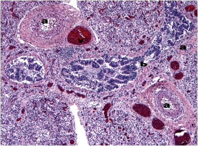
Figure 1.
Hematoxylin and eosin staining of an autopsy specimen from an infant who presented at 5 weeks demonstrates the characteristic histologic features of ACD/MPV: small pulmonary arteries with medial hyperplasia (a), congested pulmonary veins malpositioned within the same adventitial sheath (v), and a dilated bronchiole (b). Additional findings, (i.e., neutrophilic infiltrate and hemorrhage) are consequences of the hypoxemic respiratory failure and aggressive ventilatory support. (Courtesy of the Weill Cornell Medical College Department of Pathology and Laboratory Medicine.)

