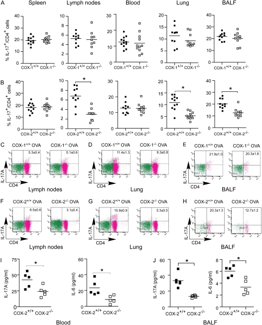
Figure 1.
Reduced Th17 cells in lung, bronchoalveolar lavage fluid (BALF), and lymph nodes of cyclooxygenase (COX)-2−/− mice after ovalbumin (OVA) sensitization and exposure in vivo. COX-1+/+, COX-1−/−, COX-2+/+, and COX-2−/− mice (n = 9–12 each) were sensitized with OVA in adjuvant. Fourteen to 21 days later, mice were exposed to inhaled OVA for 4 consecutive days. The percentages of IL-17A+ CD4+ T cells in spleen, lymph nodes, blood, lung, and BALF from COX-1+/+ versus COX-1−/− mice (A) and COX-2+/+ versus COX-2−/− mice (B) were analyzed by flow cytometry 48 hours after the last OVA exposure. Flow cytometry scattergrams show that the percentage of IL-17A+ CD4+ cells were similar in COX-1+/+ versus COX-1−/− lymph nodes (C), lung (D), and BALF (E). In contrast, COX-2−/− mice had significantly fewer IL-17A+ CD4+ cells in lymph nodes (F), lung (G), and BALF (H). IL-17A and IL-6 concentrations in blood (I) and BALF (J) were measured by ELISA and BioPlex assay 48 hours after the last OVA exposure. For panels A, B, and I, lines indicate the mean, and each symbol (solid squares, COX-1+/+ or COX-2+/+; open squares, COX-1−/− or COX-2−/−) represents an individual mouse. *P < 0.05 versus wild-type.

